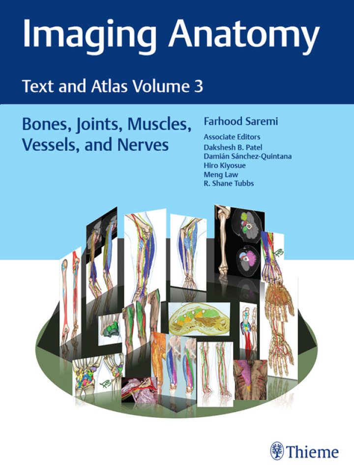
Imaging Anatomy: Text and Atlas Volume 3 – 1st Edition
Original price was: $154.99.$38.00Current price is: $38.00.
Author: Farhood Saremi; Dakshesh Patel; Damian Sanchez-Quintana; Hiro Kiyosue; Meng Law; R. Shane Tubbs
Publisher: Thieme Medical Publishers
Print ISBN: 9781626239845
Delivery Time: Within 4 hours
Copyright: 2024
500 in stock
- Save up to 60% by choosing our eBook
- High-quality PDF Format
- Lifetime & Offline Access
Imaging Anatomy: Text and Atlas Volume 3 1st Edition
Grab the definitive visual reference for modern clinical imaging: Imaging Anatomy: Text and Atlas Volume 3, 1st Edition by Farhood Saremi; Dakshesh Patel; Damian Sanchez-Quintana; Hiro Kiyosue; Meng Law; R. Shane Tubbs. This striking atlas combines precision imagery with concise, authoritative text to make anatomy immediately usable at the workstation, in the clinic, and in the classroom.
Inside you’ll find richly labeled cross-sectional images and clear anatomic diagrams that bridge textbook anatomy and real-world CT, MRI, and ultrasound appearances. Carefully organized content highlights spatial relationships, variant anatomy, and imaging landmarks so clinicians and trainees can interpret scans faster and with greater confidence. High-quality plates and side-by-side comparisons let you correlate gross anatomy with radiologic slices at a glance.
Designed for radiologists, neurologists, surgeons, residents, sonographers, and advanced students, this volume streamlines learning while supporting clinical decision-making. It’s equally valuable for exam prep and daily practice, delivering practical insights that improve diagnostic accuracy and reporting clarity. The collaborative expertise of the contributing authors ensures balanced, clinically relevant coverage informed by contemporary imaging practice worldwide.
Whether you’re working in North America, Europe, Asia, or beyond, this atlas is tailored to meet the needs of a global medical community seeking reliable, image-focused anatomy reference. Add Imaging Anatomy: Text and Atlas Volume 3 to your professional library today — a focused, visually driven resource that enhances interpretation skills and elevates patient care. Order now to bring unparalleled anatomical clarity to your imaging practice.
Note: eBooks do not include supplementary materials such as CDs, access codes, etc.