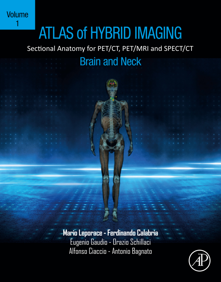
Atlas of Hybrid Imaging Sectional Anatomy for PET/CT, PET/MRI and SPECT/CT Vol. 1: Brain and Neck – 1st Edition
Original price was: $165.00.$38.00Current price is: $38.00.
Author: Mario Leporace
Publisher: Academic Press
Print ISBN: 9780323904544
Delivery Time: Within 4 hours
Copyright: 2023
500 in stock
- Save up to 60% by choosing our eBook
- High-quality PDF Format
- Lifetime & Offline Access
Atlas of Hybrid Imaging Sectional Anatomy for PET/CT, PET/MRI and SPECT/CT Vol. 1: Brain and Neck 1st Edition
Atlas of Hybrid Imaging Sectional Anatomy for PET/CT, PET/MRI and SPECT/CT Vol. 1: Brain and Neck — 1st Edition by Mario Leporace delivers an indispensable visual guide for anyone working at the intersection of anatomy and molecular imaging. This atlas grabs your attention with crisp, high-resolution sectional images that pair anatomical detail with functional PET/CT, PET/MRI and SPECT/CT correlations, making complex regions of the brain and neck immediately accessible.
Inside, you’ll find meticulously labeled transverse, coronal and sagittal slices that bridge textbook anatomy and real-world hybrid imaging. The book explains normal variants and common pathological patterns, allowing radiologists, nuclear medicine physicians, neurologists, head & neck surgeons and advanced trainees to interpret multimodality studies with confidence. Clear annotations and side-by-side comparisons speed recognition of subtle findings—critical for accurate diagnosis, staging, and treatment planning.
Practical and clinically focused, this volume emphasizes usability in busy imaging centers across Europe, North America, Asia and beyond. Whether you’re preparing for a case, teaching residents, or refining daily reporting, the atlas enhances diagnostic precision and communication between imaging teams and clinicians.
For professionals seeking a trusted reference in hybrid imaging anatomy, Mario Leporace’s atlas is an essential addition to your library. Strengthen your workflow and deepen your anatomical insight—secure your copy today to elevate practice quality and patient care in PET/CT, PET/MRI and SPECT/CT imaging.
Related Books
Medical
Writing Grant Proposals in Epidemiology, Preventive Medicine, and Biostatistics – 2nd Edition